From Wikipedia, the free encyclopedia
| Femoral artery | |
|---|---|

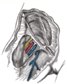
Thigh with and without the sartorius muscle, revealing the femoral artery and vein underneath |
|
| Details | |
| Source | External iliac artery |
| Branches | Superficial epigastric artery, superficial iliac circumflex, superficial external pudendal, deep external pudendal, deep femoral artery, continues as popliteal artery |
| Vein | Femoral vein |
| Supplies | Anterior compartment of thigh |
| Identifiers | |
| Latin | Arteria femoralis |
| MeSH | D005263 |
| TA98 | A12.2.16.010 |
| TA2 | 4674 |
| FMA | 70248 |
| Anatomical terminology
[edit on Wikidata] |
The femoral artery is a large artery in the thigh and the main arterial supply to the thigh and leg. The femoral artery gives off the deep femoral artery and descends along the anteromedial part of the thigh in the femoral triangle. It enters and passes through the adductor canal, and becomes the popliteal artery as it passes through the adductor hiatus in the adductor magnus near the junction of the middle and distal thirds of the thigh.[1]
The femoral artery proximal to the origin of the deep femoral artery is referred to as the common femoral artery, whereas the femoral artery distal to this origin is referred to as the superficial femoral artery.[2]
Structure[edit]

Femoral artery showing common and superficial arteries, in common usage but not listed in TA
The femoral artery represents the continuation of the external iliac artery beyond the inguinal ligament underneath which the vessel passes[2] to enter the thigh.[3] The vessel passes under the inguinal ligament just medial of the midpoint of this ligament,[2] midway between the anterior superior iliac spine and the symphysis pubis (mid-inguinal point).[citation needed]
In common usage, in clinical practice including angiology and vascular surgery, the femoral artery includes the common femoral artery, and the superficial femoral artery however, the Terminologia Anatomica (TA) only lists the femoral artery. (The TA is the international standard for human anatomical terminology developed by the Federative International Programme on Anatomical Terminology).[4]
- The common femoral artery (CFA) is located between the inferior margin of the inguinal ligament, and the branching point of the deep femoral artery. Its first three or four centimetres are enclosed, with the femoral vein, in the femoral sheath.[citation needed] In 65% of people, the common femoral artery lies anterior to the femoral vein in the upper thigh.[5] The CFA is, after the popliteal artery, the most common peripheral site of general dilatation or aneurysmal formation, at a frequency of 1/10 of the aorta.[6] Highly calcific arterial stenosis in the CFA is very difficult to treat by endovascular intervention.[7] Stent positioning in CFA may be limited by compressive or torsional forces, leading to stent fracture and/or restenosis.[7] On the other hand, lithoplasty balloon angioplasty may represent a safe tool to treat CFA stenosis.[7]
- The superficial femoral artery is the part of the femoral artery between the branching point of the deep femoral artery and the adductor hiatus, passing through the subsartorial canal.[8] The superficial femoral artery enters the adductor hiatus and becomes the popliteal artery which goes through the popliteal fossa.[9]
Relations[edit]
The relations of the femoral artery are as follows:
- Anteriorly: In the upper part of its course, it is superficial and is covered by skin and fascia. In the lower part of its course, it passes behind the sartorius muscle.
- Posteriorly: The artery lies on the psoas, which separates it from the hip joint, the pectineus, and the adductor longus. The femoral vein intervenes between the artery and the adductor longus.
- Medially: It is related to the femoral vein in the upper part of its course.
- Laterally: The femoral nerve and its branches.
Branches[edit]

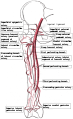
Schema of arteries of the thigh, including femoral artery and branches.
Common femoral artery
- The superficial circumflex iliac artery[10] is a small branch that runs up to the region of the anterior superior iliac spine.
- The superficial epigastric artery[10] is a small branch that crosses the inguinal ligament and runs to the region of the umbilicus.
- The superficial external pudendal artery[10] is a small branch that runs medially to supply the skin of the scrotum or labium majus as.
- The deep external pudendal artery runs medially and supplies the skin of the scrotum or labium majus.[citation needed]
- The deep femoral artery is a large and important branch that arises from the lateral side of the femoral artery about 1.5 in. (4 cm) below the inguinal ligament. It passes medially behind the femoral vessels and enters the medial fascial compartment of the thigh. It ends by becoming the fourth perforating artery. At its origin, it gives off the medial and lateral circumflex femoral arteries, and during its course it gives off three perforating arteries.[10]
Superficial femoral artery
- The descending genicular artery is a small branch that arises from the femoral artery near its termination within the adductor canal. It assists in supplying the knee joint.[citation needed]
Clinical significance[edit]
Clinical examination[edit]
The site for optimally palpating the femoral pulse is in the inner thigh, at the mid-inguinal point, halfway between the pubic symphysis and anterior superior iliac spine. Presence of a femoral pulse indicates a systolic blood pressure of more than 50 mmHg.[11]
Vascular access[edit]
Femoral artery is the frequent site of access in angiography. As the pulsation of the common femoral artery can often be palpated through the skin; and the site of maximum pulsation is used as a point of puncture for catheter access.[5] From here, wires and catheters can be directed anywhere in the arterial system for intervention or diagnostics, including the heart, brain, kidneys, arms and legs. The direction of the needle in the femoral artery can be against blood flow (retro-grade), for intervention and diagnostic towards the heart and opposite leg, or with the flow (ante-grade or ipsi-lateral) for diagnostics and intervention on the same leg. Access in either the left or right femoral artery is possible and depends on the type of intervention or diagnostic.[citation needed]
To image the lower limb vascular anatomy, the common femoral artery (CFA) is chosen as the site of entry. However, CFA entry can only be assessed by retrograde puncture. Therefore, a catheter is advanced retrogradely through the contralateral common femoral artery into common iliac artery, crossing the midline into ipsilateral CFA. The SFA can then be assessed by antegrade puncture.[12]
The femoral artery can be used to draw arterial blood when the blood pressure is so low that the radial or brachial arteries cannot be located.
Peripheral arterial disease[edit]
The femoral artery is susceptible to peripheral arterial disease.[13] When it is blocked through atherosclerosis, percutaneous intervention with access from the opposite femoral may be needed. Endarterectomy, a surgical cut down and removal of the plaque of the femoral artery is also common. If the femoral artery has to be ligated surgically to treat a popliteal aneurysm, blood can still reach the popliteal artery distal to the ligation via the genicular anastomosis. However, if flow in the femoral artery of a normal leg is suddenly disrupted, blood flow distally is rarely sufficient. The reason for this is the fact that the genicular anastomosis is only present in a minority of individuals and is always undeveloped when disease in the femoral artery is absent.[14]
See also[edit]
- Brachial artery, an arm based artery with a similar function
References[edit]
- ^ Schulte, Erik; Schumacher, Udo (2006). «Arterial Supply to the Thigh». In Ross, Lawrence M.; Lamperti, Edward D. (eds.). Thieme Atlas of Anatomy: General Anatomy and Musculoskeletal System. Thieme. p. 490. ISBN 978-3-13-142081-7.
- ^ a b c Swift, Hilary; Bordoni, Bruno (2022), «Anatomy, Bony Pelvis and Lower Limb, Femoral Artery», StatPearls, Treasure Island (FL): StatPearls Publishing, PMID 30855850, retrieved January 11, 2023
- ^ Jacob, S. (January 1, 2008), Jacob, S. (ed.), «Chapter 6 — Lower limb», Human Anatomy, Churchill Livingstone, pp. 135–179, doi:10.1016/b978-0-443-10373-5.50009-9, ISBN 978-0-443-10373-5, retrieved January 18, 2021
- ^ Kachlik D, Musil V, Blankova A, Marvanova Z, Miletin J, Trachtova D; et al. (2021). «A plea for extension of the anatomical nomenclature: Vessels». Bosn J Basic Med Sci. 21 (2): 208–220. doi:10.17305/bjbms.2020.5256. PMC 7982069. PMID 33259774.
{{cite journal}}: CS1 maint: multiple names: authors list (link) - ^ a b van den Berg, Jos C (January 2013). «Optimal Technique for Common Femoral Artery Access». Endovascular Today. Archived from the original on August 6, 2021. Retrieved August 6, 2021.
- ^ Sandgren T, Sonesson B, Ahlgren R, Länne T (1999). «The diameter of the common femoral artery in healthy human: influence of sex, age, and body size». J Vasc Surg. 29 (3): 503–10. doi:10.1016/s0741-5214(99)70279-x. PMID 10069915.
{{cite journal}}: CS1 maint: multiple names: authors list (link) - ^ a b c Trani C, Russo G, Aurigemma C, Burzotta F (2019). «The conundrum of endovascular common femoral artery treatment: a case report of lithoplasty as a viable solution». Eur Heart J Case Rep. 3 (3): ytz122. doi:10.1093/ehjcr/ytz122. PMC 6764558. PMID 31660495.
{{cite journal}}: CS1 maint: multiple names: authors list (link) - ^ Jones, Jeremy. «Femoral artery | Radiology Reference Article | Radiopaedia.org». Radiopaedia. Retrieved March 4, 2023.
- ^ Kupinski, Ann Marie (July 2019). «Venous Nomenclature». Journal of Diagnostic Medical Sonography. 35 (4): 352–353. doi:10.1177/8756479319836983. S2CID 202165116. Retrieved March 1, 2023.
- ^ a b c d Ryan, Stephanie (2011). «Chapter 8». Anatomy for diagnostic imaging (Third ed.). Elsevier Ltd. p. 306. ISBN 9780702029714.
- ^ Deakin, Charles D.; Low, J. Lorraine (September 2000). «Accuracy of the advanced trauma life support guidelines for predicting systolic blood pressure using carotid, femoral, and radial pulses: observational study». BMJ. 321 (7262): 673–4. doi:10.1136/bmj.321.7262.673. PMC 27481. PMID 10987771.
- ^ Berman, Hl; Katz, Sg; Tihansky, Dp (September 1986). «Guided direct antegrade puncture of the superficial femoral artery». American Journal of Roentgenology. 147 (3): 632–634. doi:10.2214/ajr.147.3.632. ISSN 0361-803X. PMID 2943146.
- ^ MacPherson, D. S.; Evans, D. H.; Bell, P. R. F. (January 1984). «Common femoral artery Doppler wave-forms: a comparison of three methods of objective analysis with direct pressure measurements». British Journal of Surgery. 71 (1): 46–9. doi:10.1002/bjs.1800710114. PMID 6689970. S2CID 30352039.
- ^ Sabalbal, M.; Johnson, M.; McAlister, V. (September 2013). «Absence of the genicular arterial anastomosis as generally depicted in textbooks». Annals of the Royal College of Surgeons of England. 95 (6): 405–9. doi:10.1308/003588413X13629960046831. PMC 4188287. PMID 24025288.
Additional images[edit]
-

Structures passing behind the inguinal ligament. (Femoral artery labeled at upper right.)
-

Cross-section showing structures surrounding right hip-joint.
-

Femoral sheath laid open to show its three compartments.
-

The femoral artery.
-
The spermatic cord in the inguinal canal.
-

Front of right thigh, showing surface markings for bones, femoral artery and femoral nerve.
-

Femoral artery and its major branches — right thigh, anterior view.
-

Illustration depicting main leg arteries (anterior view).
-

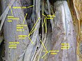
Femoral artery — deep dissection.
-
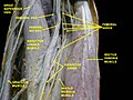
Femoral artery — deep dissection.


External links[edit]
- Anatomy photo:12:05-0101 at the SUNY Downstate Medical Center
- Cross section image: pelvis/pelvis-e12-15—Plastination Laboratory at the Medical University of Vienna
- Image at umich.edu — pulse
- Diagram at MSU Archived July 17, 2011, at the Wayback Machine
- QuantaFlo vs ABI in Peripheral Arterial Disease
На вчерашних курсах по оказанию первой помощи, которые проводит Тюменская станция скорой медицинской помощи, было не менее десяти человек. Курсы бесплатные. Все с интересом слушали врача скорой помощи Андрея Роготнева.
Полезные советы на основе практического опыта
Сразу стоит отметить, что в Интернете очень много информации по оказанию первой помощи. Курсы, которые проводят врачи скорой в Тюмени, ценны тем, что обучение проводится на основе практического опыта. Приведем простой пример. Как вы думаете, на какое время можно пережимать артерию на бедре или на плече? Многие ответят, что не более 1,5 часов. На самом деле эта рекомендация уже давно устарела, потому что через полтора часа сдавливания кровь ниже жгута свернется во всех сосудах, и конечность потом придется ампутировать. Мы ведь не на войне, и нам нужно не только спасти человеку жизнь, но еще и постараться сохранить поврежденную руку или ногу. Впрочем, обо всем по порядку.
Выясняем, откуда бежит кровь — из вены или из артерии
Перед тем, как давать полезные советы и рекомендации по остановке кровотечения, Андрей Роготнев напомнил, что кровотечения бывают венозные и артериальные. Обычные порезы мы не рассматривали, так как жизни они не угрожают. При венозном кровотечении кровь идет из вены, при артериальном — из артерии. Это в общем-то просто. Гораздо сложнее далеким от анатомии и медицины людям отличить одно кровотечение от другого. Меж тем это важно понять перед тем, как начать спасать человека. На самом деле и тут все просто: кровь из артерии идет фонтаном, но не сплошным напором, а пульсирует в такт сердцебиению, так как кровь от сердца поступает во все органы именно через артерии. По венам кровь уходит обратно, поэтому при венозном кровотечении нет такого давления, кровь бежит непрерывной струей, как вода из крана.
При артериальном кровотечении у вас в запасе не более 3-8 минут
При артериальном кровотечении вероятность гибели наиболее велика, при венозном – гибель маловероятна, опасно повреждение крупных вен на шее. В случае кровотечения из крупных артерий у окружающих есть в запасе не более 3-8 минут. За такой короткий промежуток времени скорая помощь никак не успеет доехать. Поэтому гоним прочь все страхи, надеваем на руки полиэтиленовые пакеты или резиновые перчатки, если вдруг они окажутся рядом (чтобы в случае чего не подхватить опасные вирусы), и ищем на теле точки для пальцевого прижатия артерий. Таких точек, в которых чувствуется пульсация, на теле более 10 с каждой стороны от вертикальной оси тела. Нам нужно запомнить лишь три основных: сонная, плечевая и паховая артерии.
Как пережать сонную артерию
Сонная артерия на шее — справа и слева от трахеи. В этом месте отчетливо ощущается пульс. Нащупываем и зажимаем пальцами эту артерию ниже раны. Сжимаем артерию до тех пор, пока кровь не перестанет фонтанировать. Если это произошло, вы все сделали правильно. Ни в коем случае не снижайте давление и не убирайте руку, артерию потом будет сложнее найти.
И только после того, как вам удалось остановить кровотечение, Андрей Роготнев советует делать все остальное: кричать, звать на помощь, просить прохожих вызвать скорую. Если не получилось пережать артерию пальцами, можно зафиксировать повязку (жгут, ремень или кусок ткани) через противоположную от раны руку. Таким образом, артерия будет пережата лишь с той стороны, где рана. Самый правильный вариант — сжимать сонную артерию до приезда скорой.
Как пережать плечевую артерию
Точно так же поступаем с плечевой артерией. Плечо — это часть руки от локтя до плечевого сустава. Прижмите большой палец руки к внутренней стороне плеча до кости, и вы ощутите пульсацию. Здесь и нужно зажимать артерию, обхватив руку большим и указательным пальцами, пока не подыскали, чем можно перетянуть. В аптечке у автомобилистов есть жгут, во всех остальных случаях можно воспользоваться любыми подручными средствами – шнурок из ботинок, ремень, провод зарядного устройства и т. д.
Как пережать брюшную артерию
Кровотечение из брюшной артерии (при внутреннем кровотечении) можно остановить, надавливая кулаком на живот в области пупка. Давить нужно в сторону позвоночника до тех пор, пока не остановится кровотечение либо не уменьшится. Признаки внутреннего кровотечения: человек бледный, у него учащенное сердцебиение.
Как пережать бедренную артерию
Чтобы остановить артериальное кровотечение при повреждении нижней конечности, нужно пережать кулаком паховую складку между бедром и низом живота. Здесь находится еще одна точка прижатия артерии.
Точки наложения жгута при повреждении конечностей — середина бедра или плеча
Если повреждена конечность, прижимать артерию жгутом следует выше раны. Точки наложения жгута — середина бедра и плеча. Накладывать жгут на голень или предплечье (от кисти до локтя) нет смысла, так как здесь артерии проходят между двумя костями и остановка кровотечения не гарантирована, нужно будет приложить значительное усилие, при котором можно повредить ткани.
Если вы находитесь где-то вдали от населенного пункта — в лесу или на дороге — пережимать артерию жгутом следует не более чем на 15 минут. Не более, иначе человек рискует потерять руку или ногу. Через 15 минут жгут нужно ослабить, не снимая последних два тура (оборота жгута). Отдыхаем столько же времени, сколько шли, затем снова затягиваем жгут и идем или едем дальше — туда, где нам окажут медицинскую помощь.
Как сильно нужно сжимать жгут
У многих возникает вопрос: с каким усилием нужно зажимать жгутом артерию? Жгут натягиваем до момента остановки кровотечения, затем фиксируем. Лучше попробовать самим. Усилие должно быть минимальным, как при сдавливании манжетой тонометра.
Чем воспользоваться вместо жгута
Вместо жгута можно воспользоваться тканью (шарф, рукав рубашки, штанина) или шнурок, бинт, с помощью которых можно сделать «скрутку». Охватываем бинтом конечность два раза, завязываем узел, но прижимаем не плотно к руке, оставляя расстояние с палец. Затем просовываем ключ, авторучку или любую палочку и начинаем скручивать до остановки кровотечения.
Ну вот собственно и все, что нужно знать об остановке артериального кровотечения. Главное, не упасть в обморок при виде крови. Конечно, к таким ситуациям нужно морально быть готовым заранее.
Кровь из вены при повреждении конечностей остановить гораздо проще
С венозным кровотечением все гораздо проще. Как только мы поняли, что кровь бежит из вены, зажимаем рану с помощью тугой повязки, как перевязывают обычные раны. Кровь должна остановиться.
При слабом натяжении жгута кровоток из вены только усилится
Бывают ситуации, когда люди не могут понять, бежит кровь из вены или из артерии. При этом пытаются оказать помощь. Следует помнить, что при наложении жгута кровотечение должно остановиться. При слабой фиксации жгута кровоток из вены только усилится.
Юрий Шестак, Агентство медицинской информации НЕДУГАМНЕТ
Р.Е. Калинин, И.А. Сучков, Э.А. Климентова, И.Н. Шанаев
____________________________________________________________________________________________
Рязанский государственный медицинский университет имени академика И.П. Павлова, Рязань, Российская Федерация
____________________________________________________________________________________________
Глубокая артерия бедра – крупная ветвь общей бедренной артерии, представляющая большой интерес для сосудистых и эндоваскулярных хирургов в связи с ключевой ролью в обеспечении коллатерального кровообращения между сосудами малого таза и артериями подколенно-берцового сегмента. В большинстве случаев глубокая артерия бедра отходит от заднелатеральной или задней поверхности общей бедренной артерии. В тоже время аномалии развития глубоких бедренных сосудов могут стать причиной ятрогенных повреждений при проведении открытых или эндоваскулярных вмешательств. В статье описывается клиническое наблюдение пациента, направленного на плановое ультразвуковое исследование сосудов нижних конечностей перед ангиографическим исследованием сосудов сердца и нижних конечностей, выявившее, что сразу два ствола глубокой артерии бедра имели атипичную топографию отхождения от общей бедренной артерии. Верхний ствол глубокой артерии бедра отходил от переднемедиальной поверхности общей бедренной артерии и в начальном отделе располагался над общей бедренной веной. Нижний ствол глубокой бедренной артерии отходил от переднелатеральной поверхности общей бедренной артерии. Предоперационное выявление вариантной анатомии сосудов бедренного треугольника позволило провести ангиографическое исследование коронарных сосудов через бедренную артерию на контралатеральной конечности без осложнений.
Ключевые слова: вариантная анатомия; глубокая артерия бедра; глубокая вена бедра.
2020 год, выпуск №4
Клинический случай
Читать статью (pdf) →
DOI: 10.23888/HMJ202084591-598
Для цитирования: Калинин Р.Е., Сучков И.А., Климентова Э.А., Шанаев И.Н. Редкий вариант топографии глубокой артерии бедра // Наука молодых (Eruditio Juvenium). 2020. Т. 8, №4. С. 591-598. doi:10.23888/HMJ202084591-598
Показать сокращенную информацию
| dc.contributor.author | Сенько, В. И. | |
| dc.contributor.author | Засимович, Т. В. | |
| dc.contributor.author | Павлюкевич, Е. В. | |
| dc.contributor.author | Гиль, И. В. | |
| dc.date.accessioned | 2021-02-19T09:10:31Z | |
| dc.date.available | 2021-02-19T09:10:31Z | |
| dc.date.issued | 2011 | |
| dc.identifier.citation | Вариантная анатомия бедренной артерии и её ветвей / В. И. Сенько, Т. В. Засимович, Е. В. Павлюкевич, И. В. Гиль // Весенние анатомические чтения : сборник статей научной конференции, посвященной памяти доцента З. А. Пашенко, 20 мая 2011 г. / Министерство здравоохранения Республика Беларусь, Учреждение образования «Гродненский государственный медицинский университет», Кафедра анатомии человека ; редкол.: Е. С. Околокулак (отв. ред.), Ф. Г. Гаджиева. – Гродно, 2011. – С. 40-42. | ru_RU |
| dc.identifier.isbn | 978-985-496-786-8 | |
| dc.identifier.uri | http://elib.grsmu.by/handle/files/23326 | |
| dc.language.iso | ru | ru_RU |
| dc.publisher | ГрГМУ | ru_RU |
| dc.subject | анатомия | ru_RU |
| dc.title | Вариантная анатомия бедренной артерии и её ветвей | ru_RU |
| dc.type | Article | ru_RU |
Файлы в этом документе
Данный элемент включен в следующие коллекции
- Весенние анатомические чтения : сборник статей научной конференции, посвященной памяти доцента З. А. Пашенко, 20 мая 2011 г. [21]
Показать сокращенную информацию